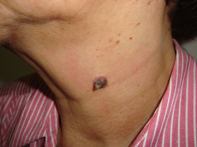
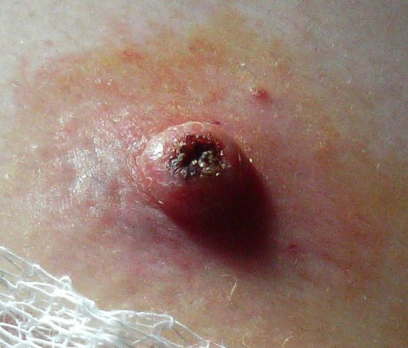
July 2012, I had this 70-year-old female patient with a skin lesion on the neck. I gave a primary clinical diagnosis of granuloma pyogenicum. My secondary clinical diagnosis is keratoacanthoma. After excision, the histopath result shows keratoacanthoma.
I read and learned from this experience.
Keratoacanthoma (KA) is classified as a skin tumor, which may be benign or malignant depending on who is talking. It is akin to squamous cell carcinoma, albeit a low-grade one. This is the reason for the conflicting views.
The defining characteristic of KA is that it is dome-shaped, symmetrical, surrounded by a smooth wall of inflamed skin, and capped with keratin scales and debris. This should be used in the clinical diagnostic process using pattern recognition.
(I will remember this and I will not forget this as I reviewed the pictures I took of the skin lesion of my patient. See below. I also looked at two pictures of published KA in the Net. This is new learning for me. I don’t think I will easily forget this because of my problem-based learning process and I spent quite some time researching and writing about it.)
From my patient.
From my patient, close-up.
From the Net.
From the Net.
***************************************
The best way to become acquainted with a subject is to write a book about it.
Benjamin Disraeli
*****************************


